透過電子顕微鏡の世界 TEM

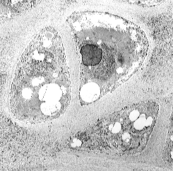
まだ、分裂して間もない軟骨細胞の姿です。
Rat meniscuses were fixed in a 2.5% glutaraldhyde in 0.1M cacodylate bufferfor 2hr, then 1% osmium tetroxide solutions, stained with uranylacetate and lead citrate (post4W.Rat-meniscus).

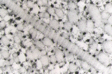
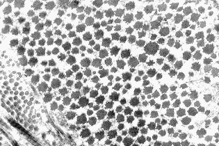
ルテニウムレッド固定したcollagen fibrilsの姿です。
collagenは約67nm周期の横紋構造をもっていますが、その周期ごとにルテニウムレッドの反応がみられます。ルテニウムレッドは強い+チャージをもっていますから、collagenの持つチャージによるものと考えられます。
Rat meniscuses were fixed with 2.5% glutaraldhyde in 1% Ruthenium Red , and then 1% osmium tetroxide in 1% Ruthenium Red solutions.

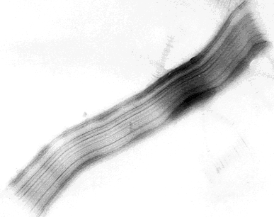
精製したcollagenをATP添加により再集合させたSLS(segment long spacing)の写真です。280nmのtropocollagen棒状分子が線維方向に対し垂直に並んでいるといわれます。
SLS was formed by dialyzing collagen solution against 0.2% ATP.2Na/0.1M acetic acid at4゜C for one day, and then stained with uranylacetate.

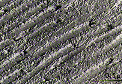
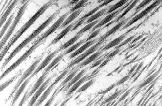
凍結割断した試料のレプリカです。
通常の透過電子顕微鏡の試料作成法では見られなかったラセン構造が表れてきます。約15nmの細線維は右巻きを示し、割断部位によっては横紋構造も見られます。
Fresh tissues were treated with 4M guanidine-HCl in buffer solution at 2 or 4 hours and fixed with 2.5% glutaraldhyde in 0.1M cacodylate buffer, glycerinated in 30% buffered glycerol for 2 or 4 hours, frozen by liqid nitrogen and fracture at -130゜C. Replication was made of platinum and carbon.


この2枚の写真は Ehlers - Danlos syndrome の症例です。
写真A は輪切りの collagen fibrils ですが、径が大きいのが特徴です。 左下に正常の collagen fibrils が見られますが、その径の大きさが比較できると思います。写真B では横紋構造も見えていますが、、、